Besides neurons, the nervous system also consists of so-called glial cells. These cells are non-excitable, and play several roles. They are generally categorized as:
Ependymal cells, which line the central cavities of the brain and the spinal column. They have microvilli and can be ciliated. The cilia of ependymal cells facilitate the flow of cerebrospinal fluid.
Choroidal epithelial cells are a slightly modified form of ependymal cells, which are found at the choroid plexus. The choroid plexus is a structure which projects into the ventricles and secretes cerebrospinal fluid into them. Choroidal epithilial cells form a tight junction, insuring that cerebrospinal fluid does not spread to the adjacent tissue.
Tanycytes are ependymal cells found at the floor of the third ventricle. These form thick processes which make contact with blood vessels and neurons, and transport hormones from the cerebrospinal fluid to capillaries and from hypothalamic neurons to the cerebrospinal fluid.
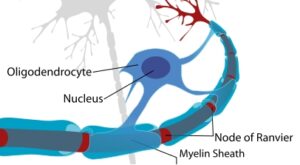
Oligodendrocites are cells which myelinate sections of neurons in the central nervous system, facilitating more rapid conduction of excitation along the axons of neurons. Oligodendrocites wrap themselves around small portions of neuronal axons, and one single Oligodendrocite can consitute several myelin segments on one or more neurons.
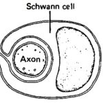
Schwann cells are responsible for myelinating axons in the peripheral nervous system, where each myelin segment on a PNS neuron will consist of one Schwann cell.
Satellite cells form a (protective?) covering around neuroganglia (neuronal groupings) such as the dorsal root ganglia and the trigeminal ganglia.
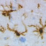
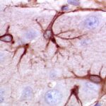
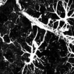
Microglia are tiny in size compared to the other types of glial cells, and are scattered throughout the brain and spinal chord. Microglia can become activated upon the even of neuronal damage, and can migrate towards the lesion site. Microglia aid in the repair of neurons by emitting specific molecules, and can also become phagocytotic, engulfing cellular debris.
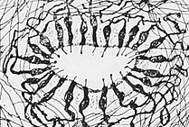
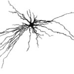
Astrocytes are possibly the most abundant type of glial cell in the nervous system, and are involved in several processes related to neurotransmitter modulation and to metabolism. There are several sub-types of astrocytes.
Protoplasmic astrocytes are found in the gray matter of the central nervous system (between layer 2 and 6 of the cerebral cortex), and play a role in the transport of nutrients from blood vessels to neurons, as well as in regulating local blood flow in the brain. Protoplasmic astrocytes also form the basis of the blood-brain barrier.
Fibrous astrocytes are primarily found in white matter tracts, and they form contacts with nerve fibres at the nodes of Ranvier. Fibrous astrocytes have the ability to react to damage to neurons by proliferating, migrating to the site of damage and by sealing off the damaged neurons, forming so-called glial scars.
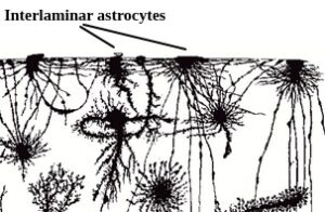
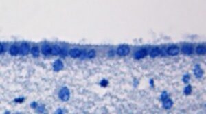
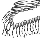
Interlaminar astrocytes and varicose astrocytes reside in cortical layer 1 (projecting into layers 3 and 4) and layers 5 and 6, respectively. The roles that interlaminar and varicose astrocytes play are currently not well known, but they are assumed to be involved in learning and cognition.