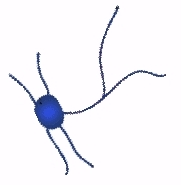
Neurons are excitable cells found throughout most of the body, and they interact with other neurons, glands, muscle tissue and the sensory apparatus. The human body contains an estimated 85 billion neurons.
Each neuron is connected to one or more (up to several hundred thousand) other neurons via junctions called synaptic terminals, which act as the main points at which neurons interact with other cells.
Neurons exhibit much greater variation than other cells in the body, and can differ from each other in their morphology, receptor types, projection patterns and electrophysiological properties.
Some classes of neurons:
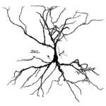
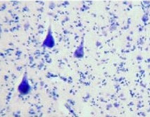
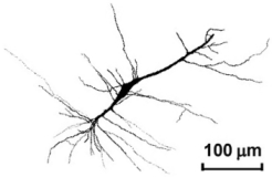
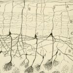
Pyramidal cells are glutamatergic (excitatory) cells with a triangle shaped cell body, and are found in the cortex, hippocampus, and amygdala. Around two thirds of neurons in the cerebral cortex are pyramidal cells. One cortical pyramidal cell can have over 10 000 connections with other neurons.
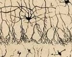
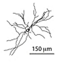
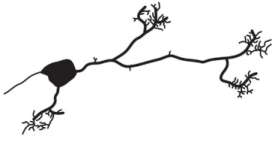
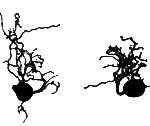
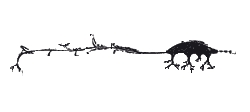
Basket cells are GABAergic (inhibitory) interneurons found in the cortex and cerebellum, which form a dense, basket-like plexus of terminals around the soma of their target cells. Basket cells appear to mediate a form of local lateral inhibition.
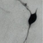
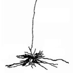
Lugaro cells are spindle-shaped inhibitory neurons located just beneath the Purkinje cell layer of the cerebellum, which appear to exert feedback inhibitory control on Purkinje cells. Lugaro cells use both GABA and glycene as neurotransmitters.
Betz cells are highly specialized large pyramidal motor neurons, and are found in areas of the primary motor cortex. These cells project all the way to the lower spinal cord, and are so large (a thickness of 70 to 100 μm) that they can be seen with the naked eye.
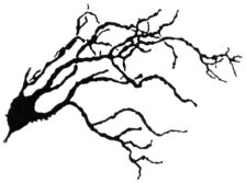
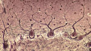
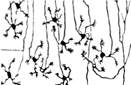
Purkinje cells are large inhibitory neurons with many branching dendrites, and are the principle neurons of the cerebellar cortex.
Medium spiny neurons are GABAergic cells, which make up over 90% of the cells of the striatum (nucleus accumbens, caudate nucleus, and putamen). Input to the striatum converges on the medium spiny neurons.
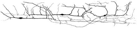
Renshaw cells are inhibitory interneurons located in the spinal cord (in lamina VII). They make inhibitory synaptic connections with several populations of motor neurons, forming a negative feedback system which may help stabilize the firing rate of the motor neurons.
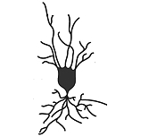
Unipolar brush cells are glutamatergic excitatory interneurons with brush-like dendrites, and are found within the granular layer of the cerebellar cortex and the granule cell domain of the cochlear nuclear complex.
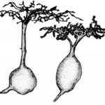
Granule cells, so-called due to their small cell bodies, are found in the cerebellum (comprising 99% of cerebellar cells), as well as in the cerebral cortex, the hippocampus, the dorsal cochlear nucleus, and the olfactory bulb. Granule cells are the smallest neurons in the brain, and are also the most numerous.
Von Economo neurons (also called spindle cells) are interneurons which connect widely separated areas of the brain. Von Economo neurons are found in the fronto-insular cortex, the anterior cingulate cortex, and the dorsolateral prefrontal cortex.
Golgi cells are large inhibitory interneurons scatterered throughout the granular layer of the cerebellum, with axons extending into the molecular layer. The majority of Golgi cells use both GABA and glycine as neurotransmitters, although a single Golgi cell will differentially mediate GABA to granule cells and glycine to unipolar brush cells.
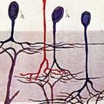
Midget cells are tiny bipolar and ganglion cells present in the retina. Midget bipolar cells receive inputs from cones, and send outputs to midget ganglion cells, the axons of which project to the parvocellular layers of the lateral geniculate nucleus of the thalamus. These cells are central to the creation of colour perception.
Parasol cells are heavily myelinated (fast conducting) ganglion cells present in the retina. Parasol ganglion cells receive activity from both rods and cones, and project axons to the magnocellular layers of the lateral geniculate nucleus of the thalamus. Parasol cells are involved in aspects of vision other than colour perception.
Photosensitive ganglion cells lie within the retina, and contain the photopigment melanopsin. These cells are critical for light entrainment, via axons to the suprachiasmatic nucleus of the hypothalamus, as well as for triggering the pupillary light reflex via projections to the pretectum in the rostral midbrain.
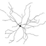
Amacrine cells are small cells found in the retina, and are so named due to their lack of an axon. Retinal amacrine cells make connections with bipolar cells and ganglion cells, and exist in a variety of subtypes.
Retinal horizontal cells are cells which receive inputs from photoreceptors and send outputs laterally to retinal bipolar cells.
Mitral cells are glutamatergic neurons in the olfactory bulb which receive inputs from receptor cells, and project to the olfactory cortex and to the limbic system.
Meynert’s cells are large pyramidal neurons present in layer V of the visual cortex, which project laterally.
Cajal-Retzius cells are a class of neurons present in the cerebral cortex during development. These glutamatergic cells are presumed to be involved in the early organization of areas of the developing cerebral cortex, and are are eliminated through cell death during early postnatal stages.
Octopus cells are glutamatergic cells present in the ventral cochlear nucleus. Octopus cells are excited by multiple auditory nerve fibers, and project to the inferior colliculus. Octopus cells may detect synchronization in the activation of groups of auditory nerve fibers, and convey its occurrence with temporal precision.